Approach Considerations
An abdominal approach is acceptable for most first- and second-trimester ultrasound examinations.
In some cases, such as those involving maternal obesity, abdominal scars due to prior surgeries, close evaluation of the cervical length during the second trimester, or gestation less than 8 weeks, a transvaginal or translabial approach may be used. The ultrasound probe used with this approach is smaller to gain access through the vagina. In addition, the sound frequency that the probe emits is higher, as the target is closer to the probe.
In general, nuchal translucency evaluation during the first trimester may be accomplished with the transabdominal approach.
First-Trimester Ultrasound
Approach to a first-trimester ultrasound
The first-trimester ultrasound examination is used mainly to confirm intrauterine pregnancy, to confirm dating, and to assess nuchal translucency. The uterus, cervix, and adnexa should be evaluated for location of a gestational sac. If a gestational sac is seen, the presence or absence of yolk sac should be reported.
Discriminatory levels of human chorionic gonadotropin (hCG) levels and ultrasound findings have been reported. Those are quantitative levels of the pregnancy hormone beta–human chorionic gonadotropin (b-hCG) and expected ultrasound findings in a viable pregnancy. They may help predict the viability of intrauterine pregnancies with uncertain viability (IPUV).
Table 1. Mean Gestational Sac Sizes at Which a Yolk Sac and an Embryo Should Be Visible (Open Table in a new window)
Visible Feature |
Mean Transvaginal Gestational Sac Diameter (mm) |
Mean Transabdominal Gestational Sac Diameter (mm) |
Serum b-hCG level (mIU/mL) |
Gestational Age (wk) |
Yolk sac visible |
8 |
20 |
7,200 |
5-6 |
Embryo visible |
16 |
25 |
10,000 |
>6 |
The yolk sac can usually be visualized if the gestational sac is approximately 1 cm in diameter. In some cases of embryonic demise, the yolk sac is deflated or irregular.
Fetal number, location, and presence and rate of heart rate should be clearly evaluated and documented. An attempt to determine chorionicity should start during the first-trimester ultrasound. Thick interfaces between gestational sacs suggest dichorionic pregnancies, and thin or absent membranes likely represent monochorionic twins.
A corpus luteum cyst may be observed in the maternal adnexa, usually 3 cm or less in diameter. Both adnexa should be evaluated for the presence of large ovarian cysts such as occurs in ovarian hyperstimulation syndrome, and solid masses such as in ovarian neoplasm. The uterus should also be evaluated for homogeneity, presence, and size and location of fibroids, especially intracavitary.
Embryonic demise
The first-trimester ultrasound presents an opportunity to identify some problematic pregnancies. Early signs of nonviable pregnancies include a gestational sac with an irregular shape or one that is not growing or b-hCG levels obtained from the patient that do not correlate with ultrasonographic findings. Embryonic demise may be diagnosed when the crown-rump length is 6 mm without fetal cardiac activity.
A study found that current guidelines regarding ultrasonographic diagnosis of miscarriage may still be associated with misdiagnoses and should be updated to take into account gestational age. [14, 15]
Ectopic pregnancy
Ectopic pregnancies or heterotopic pregnancies (one fetus inside the uterine cavity while another fetus is implanted outside the uterine cavity) may also be diagnosed. Clinically, ectopic pregnancy may manifest as pelvic pain and/or vaginal bleeding, elevated b-hCG levels, free fluid in the peritoneal cavity usually representing blood, and adnexal findings.
Anencephalia
Severe fetal anomalies can also be diagnosed in the first trimester. Anencephalic fetuses present with an absent cranium on this early ultrasound examination.
First-trimester sonographic markers for aneuploidy
Early tests for aneuploidy include the measurement of the skin swelling behind the fetal neck (the nuchal translucency), along with two biochemical markers, pregnancy-associated plasma protein A (PAPP-A) and free-hCG. First-trimester screening has a detection rate of 87% for aneuploidy. Other anatomical markers for aneuploidy on the first-trimester ultrasound include the absence of nasal bone, increased frontomaxillary angle measurement based on gestational age, the appearance of an objective decrease in blood flow during an atrial contraction (a-wave abnormalities) on ductus venosus Doppler evaluation, and increased tricuspid valve regurgitation.
However, not all of these markers can currently be assessed in the general population. [16]
Second- and Third-Trimester Ultrasound Evaluation
Approach to the second-trimester obstetric ultrasound
The most common indication for a second-trimester ultrasound examination is evaluation of fetal anatomy, usually between 18 and 20 weeks’ gestation. At this point, the embryologic period is well passed, and fetal organs are, for the most part, easily visualized and evaluated. Fetal position, early pregnancy, anterior placenta, multiple pregnancies, uterine fibroids, and maternal obesity may impede a good anatomical evaluation of the fetus at this stage. If the examination is inconclusive because of one of these factors, the patient may be asked to return to finalize the fetal anatomical evaluation at a later date.
In some cases, owing to choice or lack of early access to prenatal care, patients are unable to obtain an early ultrasound examination to confirm the gestational age of the pregnancy, number of fetuses, and chorionicity, which can be determined with a second-trimester ultrasound.
While a first-trimester ultrasound is considered a better tool for gestational dating, the second-trimester ultrasound can be used to determine gestational age. A composite of multiple fetal measures is made and averaged. The common practice is not to override dates based on the calculation of last menstrual period unless there is a difference of 5 days between dates determined by last menstrual period and first-trimester ultrasound or a difference of 10 days between the last menstrual period and second-trimester ultrasound.
Multiple pregnancies
Evaluation of fetal chorionicity when multiples are present may be performed similarly as on the first-trimester ultrasound. If the fetuses are located in different gestational sacs and the membrane dividing the fetuses is thick and has a broad base, the pregnancy is more likely dichorionic. Conversely, if the membrane dividing the twins is thin without a broad base (called a “twin peak” or “delta sign”) or absent, it suggests a monochorionic pregnancy. If the fetuses are different sexes, dichorionic pregnancy is diagnosed.
Placental evaluation
If the patient presents with acute vaginal bleeding, ultrasound may help determine cause. Placenta previa occurs when part or the entire placenta covers the internal cervical os. This diagnosis is important, as it requires cesarean delivery. Abruptio placentae refers to early separation of part or the complete placenta. A hypoechoic area may be seen on the interface between the uterus and the placenta. Vasa previa is rarer and is diagnosed by visualizing placental blood vessels running across the internal cervical os.
Fetal ultrasound testing
Fetal well-being may be evaluated with various measures. A biophysical profile (BPP) is composed of a series of measurements to assess fetal hypoxia. They include amniotic fluid evaluation, breathing movement, gross body movements, and fine movement. A scoring system consisting of either 0 or 2 points is then applied to the fetal evaluation.
Measurement of the fetal umbilical artery (UA) Doppler indicates the resistance that fetal blood flow finds at the placental level. To obtain fetal UA Doppler, the angle of insonation should be as close to zero degrees to the umbilical artery as possible. A comparison of forward blood flow between fetal systole and diastole is obtained. A large systolic-to-diastolic ratio indicates higher resistance of blood flow at the placental level.
Maternal anatomy to be evaluated includes the uterus, adnexa, and cervix.
Fetal structures to be evaluated include the head, face, and neck, including the cerebellum, choroid plexus, cisterna magna, lateral cerebral ventricles, midline falx, cavum septi pellucidi, and upper lip.
Most of the fetal syndromes, aneuploidies, and severe fetal anomalies manifest as fetal CNS anomalies. A normal ultrasound finding does not completely rule out fetal aneuploidies but is reassuring. If the patient had a prior pregnancy complicated by CNS anomalies, the risk of recurrence in a later pregnancy is about 2%.
Fetal growth
Similarly, by following the same parameters measured to obtain gestational age on the second-trimester ultrasound, the pregnancy may be monitored for objective estimation of fetal growth. Clinically, fetal growth is monitored by measuring the fundal height during routine obstetric visits. Fetal measurements obtained to determine fetal age and weight include biparietal diameter, head circumference, abdominal circumference, and femur and humerus length. When growth of fundal height is not as expected in some twin pregnancies or others pregnancies at risk for growth restriction, serial second- and third-trimester ultrasounds may be performed to evaluate adequate fetal growth. Because of variability in ultrasound measurements, a 3-week wait between growth ultrasounds is used to determine fetal growth.
Fetal head circumference should be measured in a plane that includes the cavum septum pellucidum, falx cerebri, and thalamus. To measure the biparietal diameter (BPD), the electronic calipers should be placed from the outside the calvarium proximally to the inside of the calvarium distally, crossing the falx cerebri at a 90° angle.
The same anatomical landmarks used to measure the biparietal diameter are used to measure the fetal head circumference. This measurement is obtained by measuring the outer circumference formed by the calvarium. Head measurements may be helpful to indicate fetal age, microcephaly, or macrocephaly.
Objectively, the cephalic index (CI) is defined as the width of the head divided by the length in a percentage scale. The normal value is between 70 and 80, but an abnormal value does not necessarily indicate pathology. If the cephalic index is more than 80, the fetal head will have a rounded appearance, also called brachycephalia. If the cephalic index is less than 70, the head may appear flat, also known as dolichocephalia. Minor changes in the shape of the fetal head are normal. However, brachycephalia may be seen in trisomy 18, and dolichocephalia may be seen is preterm babies or craniosynostosis.
Fetal anatomy: CNS
The cerebellum is located in the posterior inferior aspect of the brain. It has a large mass of cerebral cortex above and the Pons below. It is divided in two hemispheres. The cerebellar surface folds into itself, increasing the area. Those folds are called lobules. There are 3 major areas: the flocculonodular, anterior, and posterior lobes. This organ has more neurons than the rest of the brain. The functions are related to gait, language, and body tone. Fetal evaluation of the cerebellum includes its presence, shape, and size. During a second-trimester ultrasound, cerebellum is measured by the outer length of the posterior cerebellar hemispheres, at the same plane that the septum cavum pellucidum, cisterna magna, and nuchal fold are seen.
Posterior fossa deformities are seen with Chiari II malformations (also called Arnold-Chiari), in which parts of the cerebellum and brainstem are pulled downward into the foramen ovale owing to a spinal defect. The sonogram shows obliteration of the posterior fossa structures, usually with elongation of the cisterna magna (banana sign) and flattening of the frontal cephalic bones (lemon sign).
The cisterna magna is also located in the posterior fossa, posterior to the cerebellum. It is part of the openings of the subarachnoid space. Dilatation of the cisterna magna may occur in conditions such as Dandy-Walker malformation, in which there is an abnormal communication between the fourth ventricle and the cisterna magna via a defect in the cerebellar vermis.
The human brain has 4 choroid plexus. They are built as a filter between the blood and the cerebrospinal fluid (CSF) and consist of many blood vessels separated from the ventricular space by choroid epithelial cells that, by passive and active transport, maintain the balance of the cerebrospinal fluid. They are located in the superior horn of the lateral ventricles. On ultrasound, they appear as a bright half-moon–shaped echo area in the ventricle. Choroid plexus cysts are found in 1 per 100 fetuses with trisomy 18; however, if the only abnormal finding is a choroid plexus cyst, the risk for trisomy 18 is 1 in 477.
The American College of Obstetricians and Gynecologists (ACOG) recommends considering amniocentesis if there are other “soft” markers for aneuploidy, if the blood markers for aneuploidy are abnormal, or if the mother will be aged 32 years at the time of term delivery. A choroid plexus that is floating in the lateral ventricle is called a floating or “drooping” choroid, which is caused by an enlarged lateral ventricle.
The ventricular system of the brain is composed of the lateral ventricles and the third and the fourth ventricles. Cerebrospinal fluid (CSF) is produced by blood filtered by ependymal cells on the choroid plexus. CSF travels along all ventricles to be reabsorbed through the arachnoid villi.
During second-trimester ultrasound, the lateral ventricles are measured in a plane where the cavum septum pellucidum is visible anteriorly, from the internal membranes of the lateral ventricles just behind the choroid plexus. A blockage from the reabsorption system may cause an enlarged lateral ventricle, with increased CSF pressure, which is called ventriculomegaly, a condition seen with hydrocephalus. If the cause of the hydrocephalus is not evident, it is called simple hydrocephalus. In some cases, hydrocephalus may occur along with other conditions such as Dandy-Walker malformation or agenesis of the corpus callosum.
The cavum septum pellucidum is composed of two different layers of gray and white matter. The space between the two layers is the cavum, which disappears during adult life. It is closely related to the corpus callosum. Absence of the cavum on a second-trimester ultrasound may indicate absence of the corpus callosum, which may be associated with multiple syndromes, including trisomy 13 or 18 or Dandy-Walker malformation. Agenesis of the corpus callosum may be confirmed with ultrasound of the midcoronal planes.
Fetal anatomy: Spine
The spinal evaluation of the fetus begins with an anatomic assessment of the head, abnormalities of which reflect some spinal problems. For instance, Chiari II malformations are often seen in fetuses with spina bifida. Owing to the decreased pressure in the spine, contents of the posterior fossa herniate through the foramen magnum into the spinal canal. There is displacement of the cerebellar vermis, fourth ventricle, and medulla oblongata. The cistern magna enlarges, becoming banana-shaped. The frontal bones collapse and the head may take on a lemon shape.
The most common spinal anomalies are the ones associated with spina bifida, in which failure of the vertebral bones to close allows part of the spinal cord to lie outside the neural canal. In cases of spina bifida occulta, the vertebral schisis is covered with skin, and it may appear as a lipoma or dimple at birth. If the defect is open, it is referred as a spina bifida aperta, and it is further classified as a meningocele (if it is covered by a thin meningeal membrane) or myelomeningocele (if the sac contains neural tissue).
The International Society of Ultrasound in Obstetrics and Gynecology (ISUOG) recommends that, when possible, a longitudinal section of the fetal spine should be obtained to screen for open and closed spinal dysraphism. Targeted fetal neurosonography is recommended if there is suspicion of a brain or spinal abnormality during the obstetric ultrasound examination. [17]
Fetal anatomy: Chest
The fetal chest evaluation includes the fetal heart and lungs. The chest view should include a 4-chamber heart and outflow tracts, if possible. A detailed cardiac ultrasound is beyond the scope of this article and is reserved for patients with a prior history of a fetus with cardiac anomalies, patients with overt diabetes, suspicions of a syndrome that affects the fetal heart, and patients with abnormally appearing fetal cardiac anatomy based on second-trimester ultrasound.
In the evaluation of the fetal heart, the position of the fetal heart in relation to the fetal body should be assessed first. The fetal stomach and the apex of the fetal heart should be on the left side of the fetus.
Next, a 4-chamber view of the fetal heart should be obtained. The transverse view of the fetal heart is obtained right above the fetal diaphragm. With minor adjustments, a 4-chamber view is acquired. The apex should be pointing to the left side, and the right ventricle should be the most anterior chamber. The upper chambers should be equal in size to the lower chambers, and both sides should be of similar size during both systole and diastole. This view allows the sonographer to evaluate the interventricular septum. The size of the heart should be about half the size of the whole thorax. [18]
Movement evaluation during this view includes free movement of both atrioventricular valves, as well as the foramen ovale flap in the left atrium. In some cases, an echogenic intracardiac focus that represents calcifications on the papillary muscle is seen and is a marker for fetal aneuploidy. With this view, more than half of the congenital cardiac anomalies may be detected, including those related to cardiac position, cardiac septa, cardiac chambers, and masses. Left outflow and right outflow views may be obtained, if possible.
Additional cardiac views may be obtained for a more complete fetal cardiac evaluation. These include the long axis of the left ventricle, short axis of the great vessels, aortic arch, and pulmonary artery view.
Fetal heart tones may be visible as early as 5 weeks’ gestation. The 5 components of the fetal heart motion to be evaluated include rate, rhythm, atrioventricular association, structural anomalies, and evaluation for hydrops. The fetal heart rate may change during the course of the pregnancy but should generally be between 110 and 180 beats per minute. Routinely, an M-mode picture of the fetal heart is taken. Fetal heart rhythm may be constant or variable; minor variability is normal, but significant rate variability should be noted, and, if the fetus is viable, the obstetrician should be notified immediately.
The fetal diaphragm is evaluated for continuity. Congenital diaphragmatic hernia is the most common anomaly in the fetal thorax if cardiac problems are excluded. They are difficult to observe with ultrasound tend to grow as the pregnancy progresses. The diagnosis may be made upon observation of loops of bowel in the thorax or discontinuity of the diaphragm muscle.
Cysts in the region of the lungs may result from cystic adenomatous hyperplasia or bronchopulmonary sequestration. There is hamartomatous involvement in cystic adenomatous hyperplasia, and the prognosis depends on the size and type. In bronchopulmonary sequestration, a portion of the bronchopulmonary system develops separately and is seen as a hyperechoic area at the lung base.
Fetal anatomy: Abdomen
Components of the abdominal-area evaluation should include presence, size, and position and of the stomach, kidneys, bladder, umbilical cord insertion site into the fetal abdomen, and umbilical cord vessels.
The plane for abdominal circumference should include the outer margins of a transverse plane that includes the fetal stomach, spine, liver, and the umbilical portion of the left portal vein equidistant from both sides of the abdomen. A small circumference may indicate a growth-restricted fetus. The fetal liver can be evaluated for size and presence of masses or cysts. Liver calcifications have been described as part of fetal viral infections. A “double-bubble” stomach indicates duodenal atresia, a marker for Down syndrome. Absence of the fetal stomach may be related to other fetal anomalies.
Esophageal atresia may be diagnosed with ultrasound based on the absence of a visible fetal stomach and the presence of polyhydramnios; however, the diagnostic yield with ultrasound is only about 50%. Bowel dilatation may indicate large-bowel obstruction or anal atresia. Hyperechoic bowel is seen in cases of meconium, bleeding, or cystic fibrosis.
Two anterior abdominal defects are worth mentioning. Omphalocele is a midline defect covered by a membrane. If the defect is large enough, it may include the fetal liver. The association with fetal syndromes is high. Gastroschisis is a lateral defect to the umbilical cord, not covered by a membrane. On ultrasound, it is visualized as free loops of bowel in the amniotic sac. It has been associated with some fetal syndromes, but at a lower rate than omphalocele.
Fetal kidneys are located on both sides of the fetal spine, with an ovoid shape in a longitudinal view and a round shape in a transverse view. Fetal kidneys enlarge with gestational age; therefore, they are easier to visualize as pregnancy progresses and the fetus grows. It is not routinely necessary to measure the kidneys. Care should be taken in not confuse the fetal adrenal gland as part of the kidneys, as it may be larger than in adults. Minor fluid collection in the kidneys is normal, but more than 4 mm before 32 weeks’ gestation or more than 7 mm after 32 weeks’ gestation fluid collection or pyelectasis may be a marker of aneuploidy.
In fetuses who present with obstruction of the renal outlet, the affected kidney is visualized on ultrasound as a paraspinal sac, cysts, or abnormal ureteral dilatations. Absence of kidneys is rarer but carries a lethal prognosis. Polycystic kidneys are a bilateral disorder representing tubular ectasia, usually appearing after the second trimester. Meckel-Gruber syndrome is characterized by polycystic kidneys with encephalocele and polydactyly.
Fetal bladder is seen as a hypoechoic area caudally on the fetus. The filling cycle of the bladder is 60-90 minutes; therefore if an initial view of the fetus does not demonstrate a bladder, repeat imaging is recommended. In rare cases such as in urinary obstruction, the identification of fetal sex is medically indicated.
The umbilical cord may be evaluated by applying color Doppler interrogation to the area around the bladder and visualizing the two umbilical arteries around the bladder. Care should be taken to obtain a transverse view of the fetal abdomen and not to confuse the fetal iliac arteries.
Fetal anatomy: Extremities
The presence or absence of fetal extremities should also be documented. This is done by demonstrating one bone in the proximal extremity and two bones distally. Measurement of the long bones is obtained to evaluate fetal growth.
A single-center study by Dicke et al indicated that although ultrasound examination allows most cases of abnormal hand position, limb reduction defect, and arthrogryposis to be detected prenatally, at least 20-25% of these abnormalities will probably be missed. The sensitivity of ultrasound was found to be as follows [19] :
-
Polydactyly: 19.1%
-
Abnormal hand position: 76.0%
-
Limb reduction defect involving the long bones: 76.1%
-
Arthrogryposis: 81.3%
-
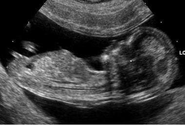
Embryo at 12 weeks' gestation.
-
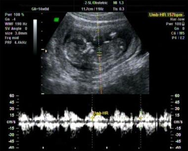
Fetal heartbeat at 20 weeks' gestation.
Tables
Gestational age in weeks (by LMP) |
Discrepancy between menstrual dating and ultrasound estimates |
8 6/7 or less |
> 5 days |
9 0/7 to 13 6/7 |
> 7 days |
14 0/7 to 15 6/7 |
> 7 days |
16 0/7 to 21 6/7 |
> 10 days |
22 0/7 to 27 6/7 |
> 14 days |
28 0/7 and beyond |
> 21 days |
Visible Feature |
Mean Transvaginal Gestational Sac Diameter (mm) |
Mean Transabdominal Gestational Sac Diameter (mm) |
Serum b-hCG level (mIU/mL) |
Gestational Age (wk) |
Yolk sac visible |
8 |
20 |
7,200 |
5-6 |
Embryo visible |
16 |
25 |
10,000 |
>6 |


