Applebaum H, Sydorak R. Cystic abdominal disease: mesenteric, omental, solid organ. Ziegler MM, Azizkhan RG, von Allmen D, Weber TR, ed. Operative Pediatric Surgery. 2nd ed. New York: McGraw-Hill; 2014. 664-8.
Ricketts RR. Mesenteric and omental cysts. Coran AG, Adzick NS, Krummel TM, Laberge J-M, Shamberger RC, Caldamone AA, eds. Pediatric Surgery. 7th ed. Philadelphia: Elsevier Saunders; 2012. Vol 2: 1165-70.
Tripathy PK, Jena PK, Pattnaik K. Management outcomes of mesenteric cysts in paediatric age group. Afr J Paediatr Surg. 2022 Jan-Mar. 19 (1):32-35. [QxMD MEDLINE Link]. [Full Text].
Egozi EI, Ricketts RR. Mesenteric and omental cysts in children. Am Surg. 1997 Mar. 63 (3):287-90. [QxMD MEDLINE Link].
Gross RE. The Surgery of Infancy and Childhood. Philadelphia: WB Saunders; 1953. 377-83.
Bliss DP Jr, Coffin CM, Bower RJ, Stockmann PT, Ternberg JL. Mesenteric cysts in children. Surgery. 1994 May. 115 (5):571-7. [QxMD MEDLINE Link].
Skandalakis JE, Gray SW, Ricketts RR. The lymphatic system. Skandalakis JE, Gray SW, eds. Embryology for Surgeons. 2nd ed. Baltimore: Lippincott Williams & Wilkins; 1994. 891-7.
Lee DL, Madhuvrata P, Reed MW, Balasubramanian SP. Chylous mesenteric cyst: A diagnostic dilemma. Asian J Surg. 2016 Jul. 39 (3):182-6. [QxMD MEDLINE Link].
Kushwaha JK, Gupta R, Mohanti S, Kumar S. Primary mesenteric hydatid cyst. BMJ Case Rep. 2012 Jul 9. 2012:[QxMD MEDLINE Link].
Aytekin S, Alyamac G. Two new cases with Costello syndrome. Dermatol Online J. 2013 Aug 15. 19 (8):19267. [QxMD MEDLINE Link].
Kurtz RJ, Heimann TM, Holt J, Beck AR. Mesenteric and retroperitoneal cysts. Ann Surg. 1986 Jan. 203 (1):109-12. [QxMD MEDLINE Link]. [Full Text].
Gupta RK, Sah S, Sah PL, Shah BP. Congenital omental cyst. BMJ Case Rep. 2012 Aug 2. 2012:[QxMD MEDLINE Link].
Schols RM, Stassen LP, Keymeulen KB, Bouvy ND. Dermoid cyst of the greater omentum: rare and innocent?. BMJ Case Rep. 2013 Feb 28. 2013:[QxMD MEDLINE Link].
Sforza M, Andjelkov K, Ivanov D, Maricić Z, Krstić S. A rare case of benign omentum teratoma. Srp Arh Celok Lek. 2012 May-Jun. 140 (5-6):362-4. [QxMD MEDLINE Link].
Chung MA, Brandt ML, St-Vil D, Yazbeck S. Mesenteric cysts in children. J Pediatr Surg. 1991 Nov. 26 (11):1306-8. [QxMD MEDLINE Link].
Hebra A, Brown MF, McGeehin KM, Ross AJ 3rd. Mesenteric, omental, and retroperitoneal cysts in children: a clinical study of 22 cases. South Med J. 1993 Feb. 86 (2):173-6. [QxMD MEDLINE Link].
Kosir MA, Sonnino RE, Gauderer MW. Pediatric abdominal lymphangiomas: a plea for early recognition. J Pediatr Surg. 1991 Nov. 26 (11):1309-13. [QxMD MEDLINE Link].
Alemu H, Alemu S, Berhane M. Omental Cyst Presenting as an Acute Abdomen in a Pediatric Patient: A Case Report. Int Med Case Rep J. 2022. 15:43-46. [QxMD MEDLINE Link]. [Full Text].
Robbins KJ, Antiel RM, Shakhsheer BA. Omental cyst: a case report and review of the literature. Ann Pediatr Surg. 2021. 17 (1):62. [QxMD MEDLINE Link]. [Full Text].
Lockhart C, Kennedy A, Ali S, McManus D, Johnston SD. Mesenteric cysts: a rare cause of abdominal pain. Ulster Med J. 2005 May. 74 (1):60-2. [QxMD MEDLINE Link].
Yoon JW, Choi DY, Oh YK, Lee SH, Gang DB, Yu ST. A Case of Mesenteric Cyst in a 4-Year-Old Child with Acute Abdominal Pain. Pediatr Gastroenterol Hepatol Nutr. 2017 Dec. 20 (4):268-272. [QxMD MEDLINE Link]. [Full Text].
Prasad KK, Jain M, Gupta RK. Omental cyst in children presenting as pseudoascites: report of two cases and review of the literature. Indian J Pathol Microbiol. 2001 Apr. 44 (2):153-5. [QxMD MEDLINE Link].
Karhan AN, Soyer T, Gunes A, Talim B, Karnak I, Oguz B, et al. Giant Omental Cyst (Lymphangioma) Mimicking Ascites and Tuberculosis. Iran J Radiol. 2016 Jul. 13 (3):e31943. [QxMD MEDLINE Link]. [Full Text].
Makhija D, Shah H, Tiwari C, Jayaswal S, Khedkar K, Waghmare M. Mesenteric cyst(s) presenting as acute intestinal obstruction in children: Three cases and literature review. Int J Pediatr Adolesc Med. 2016 Sep. 3 (3):109-111. [QxMD MEDLINE Link]. [Full Text].
Maung M, Saing H. Intestinal volvulus: an experience in a developing country. J Pediatr Surg. 1995 May. 30 (5):679-81. [QxMD MEDLINE Link].
Mohanty SK, Bal RK, Maudar KK. Mesenteric cyst--an unusual presentation. J Pediatr Surg. 1998 May. 33 (5):792-3. [QxMD MEDLINE Link].
Monabati A, Safavi M, Solhjoo F. Extragastrointestinal Stromal Tumor Presenting as Omental Cyst. J Gastrointest Surg. 2016 Jun. 20 (6):1275-7. [QxMD MEDLINE Link].
Kokhanovsky N, Nachtigal A, Reindorp N, Shinhar D, Zeina AR. Giant omental hemorrhagic cyst presenting as acute hemorrhagic anemia in a 21-month-old infant. Pediatr Emerg Care. 2014 Mar. 30 (3):188-90. [QxMD MEDLINE Link].
Chou YH, Tiu CM, Lui WY, Chang T. Mesenteric and omental cysts: an ultrasonographic and clinical study of 15 patients. Gastrointest Radiol. 1991 Fall. 16 (4):311-4. [QxMD MEDLINE Link].
Wootton-Gorges SL, Thomas KB, Harned RK, Wu SR, Stein-Wexler R, Strain JD. Giant cystic abdominal masses in children. Pediatr Radiol. 2005 Dec. 35 (12):1277-88. [QxMD MEDLINE Link].
Nakano T, Kobayashi M, Usui T, Hanazaki K. Omental pseudocyst. Radiat Med. 2007 Aug 1. 25 (7):364-7. [QxMD MEDLINE Link].
Luo CC, Huang CS, Chao HC, Chu SM, Hsueh C. Intra-abdominal cystic lymphangiomas in infancy and childhood. Chang Gung Med J. 2004 Jul. 27 (7):509-14. [QxMD MEDLINE Link].
Alqahtani A, Nguyen LT, Flageole H, Shaw K, Laberge JM. 25 years' experience with lymphangiomas in children. J Pediatr Surg. 1999 Jul. 34 (7):1164-8. [QxMD MEDLINE Link].
Catania VD, Briganti V, Di Giacomo V, Miele V, Signore F, de Waure C, et al. Fetal intra-abdominal cysts: accuracy and predictive value of prenatal ultrasound. J Matern Fetal Neonatal Med. 2016. 29 (10):1691-9. [QxMD MEDLINE Link].
Cass DL. Fetal abdominal tumors and cysts. Transl Pediatr. 2021 May. 10 (5):1530-1541. [QxMD MEDLINE Link]. [Full Text].
Rezaee-Azandaryani A, Ghorbanpour M, Taghipour M, Yamini A. A Case Report of a Huge Mesenteric Cyst in a 5-Year-Old Girl: A Rare and Challenging Finding in Radiological Assessment. Adv J Emerg Med. 2020 Spring. 4 (2):e31. [QxMD MEDLINE Link]. [Full Text].
Chen Q, Zhang S, Luo W, Cai D, Zhang Y, Huang Z, et al. Robotic-assisted laparoscopic management of mesenteric cysts in children. Front Pediatr. 2022. 10:1089168. [QxMD MEDLINE Link]. [Full Text].
Polat C, Yilmaz S, Arikan Y, Mahallesi D, Caddesi KM. Mesenteric cysts. Surg Endosc. 2004 Jan. 18 (1):169. [QxMD MEDLINE Link].
Trompetas V, Varsamidakis N. Laparoscopic management of mesenteric cysts. Surg Endosc. 2003 Dec. 17 (12):2036. [QxMD MEDLINE Link].
Bhandarwar AH, Tayade MB, Borisa AD, Kasat GV. Laparoscopic excision of mesenteric cyst of sigmoid mesocolon. J Minim Access Surg. 2013 Jan. 9 (1):37-9. [QxMD MEDLINE Link]. [Full Text].
Pampal A, Yagmurlu A. Successful laparoscopic removal of mesenteric and omental cysts in toddlers: 3 cases with a literature review. J Pediatr Surg. 2012 Aug. 47 (8):e5-8. [QxMD MEDLINE Link].
Gałązka P, Redloch K, Kroczek K, Styczyński J. Minimally Invasive Surgery for Congenital Abdominal Cystic Lesions in Newborns and Infants. In Vivo. 2020 May-Jun. 34 (3):1215-1221. [QxMD MEDLINE Link].
Al-Harfoushi R, Stevenson L, Binnie N. Mesenteric cyst: drained and marsupialised laparoscopically avoiding enterectomy. BMJ Case Rep. 2012 Jul 3. 2012:[QxMD MEDLINE Link].
Ma A, Ayre K, Wijeyekoon S. Giant mesenteric cyst: a rare cause of abdominal distension diagnosed with CT and managed with ultrasound-guided drainage. BMJ Case Rep. 2012 Sep 3. 2012:[QxMD MEDLINE Link].
Chang TS, Ricketts R, Abramowsky CR, Cotter BD, Steelman CK, Husain A, et al. Mesenteric cystic masses: a series of 21 pediatric cases and review of the literature. Fetal Pediatr Pathol. 2011. 30 (1):40-4. [QxMD MEDLINE Link].
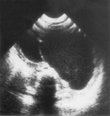 Ultrasound image demonstrating a thin-walled mesenteric cyst with an internal septum.


