Pediatric Mesenteric Cyst

  • Etiology: mesothelial-lined peritoneal surfaces failing to coalesce, arises from root of mesentery
  • US: thin walled simple cyst with posterior acoustic enhancement

Radiology Cases of Mesenteric Cyst

CT of mesenteric cyst
Axial CT with contrast of the abdomen shows a large simple cystic mass in the left side of the abdomen.
CT of mesenteric cyst
Image from a small bowel follow through exam (above) shows the small bowel to be entirely opacified and to be elevated out of the pelvis with the exception of a single loop in the middle of the pelvis. Axial CT without intravenous and with oral contrast of the abdomen (below) shows a large low density multiseptated mass in the pelvis.

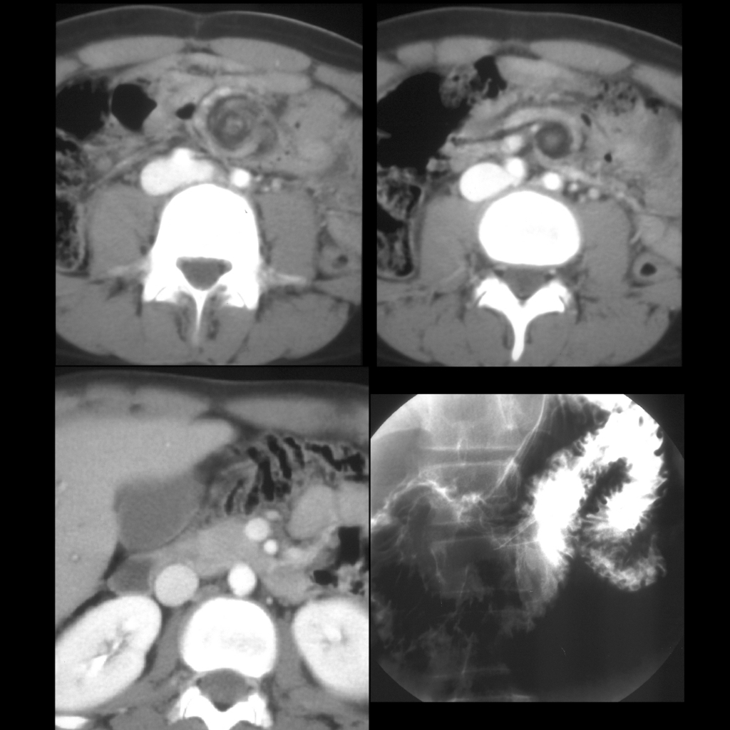
Radiology Case of Mesenteric Cyst Causing Small Bowel Volvulus

CT of small bowel volvulus around mesenteric cyst
Two contiguous axial images from a CT with contrast of the abdomen show a whirlpool sign of swirling of the mesenteric vessels (above left) around a round low density structure just to the left of the spine (above right). Another axial image from the same exam (below left) shows a normal relationship of the superior mesenteric vein to the right of the superior mesenteric artery although both appear to be shifted to the left. AP image from a subsequent upper GI (below right) shows normal position of the ligament of Treitz in the left upper quadrant.

Surgery Cases of Mesenteric Cyst

Surgical image shows a large thin-walled cystic structure arising from the mesentery of the small bowel.