
Transvaginal Ultrasound in the Diagnosis and Assessment of Endometriosis—An Overview: How, Why, and When

 ,
,  ,
,  and
and 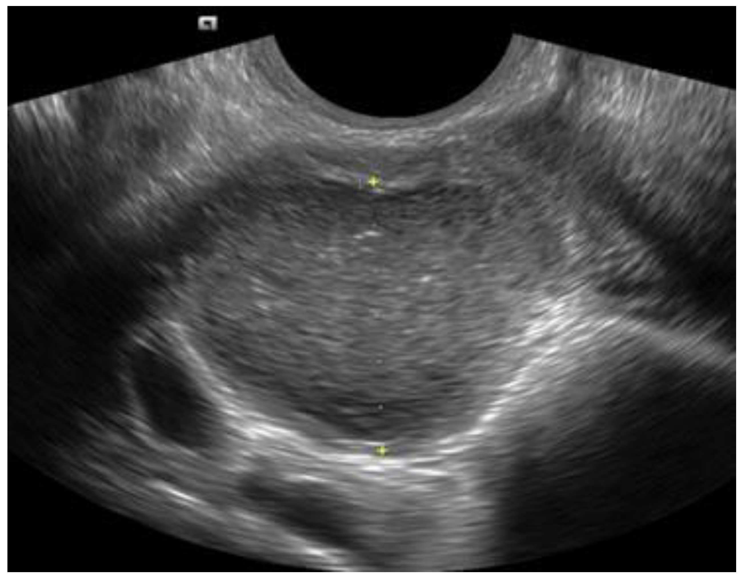
Abstract
:1. Introduction
2. Results and Discussion
2.1. Ovarian Endometrioma (OΜA)
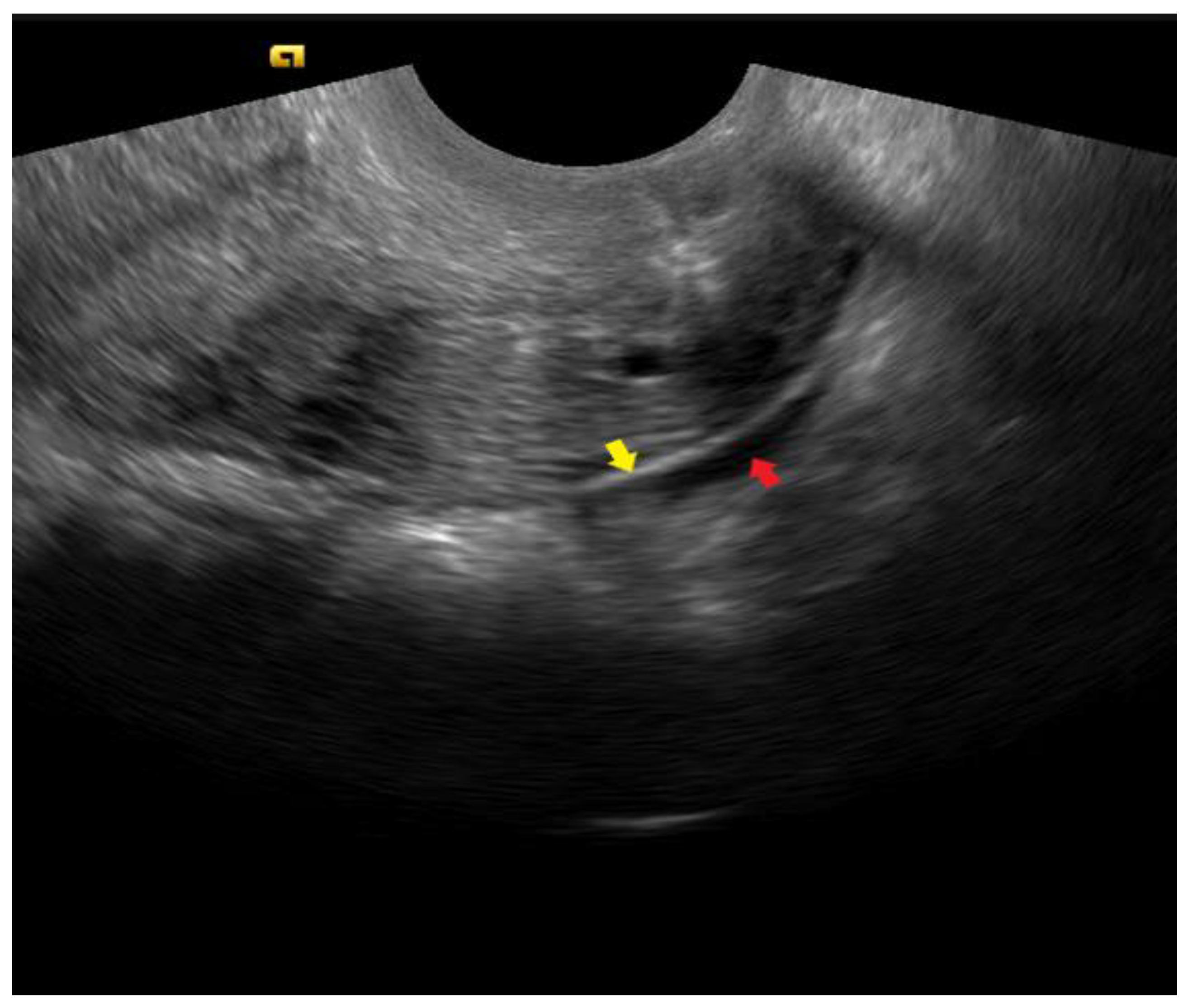
2.2. Superficial Peritoneal Endometriosis (SUP)
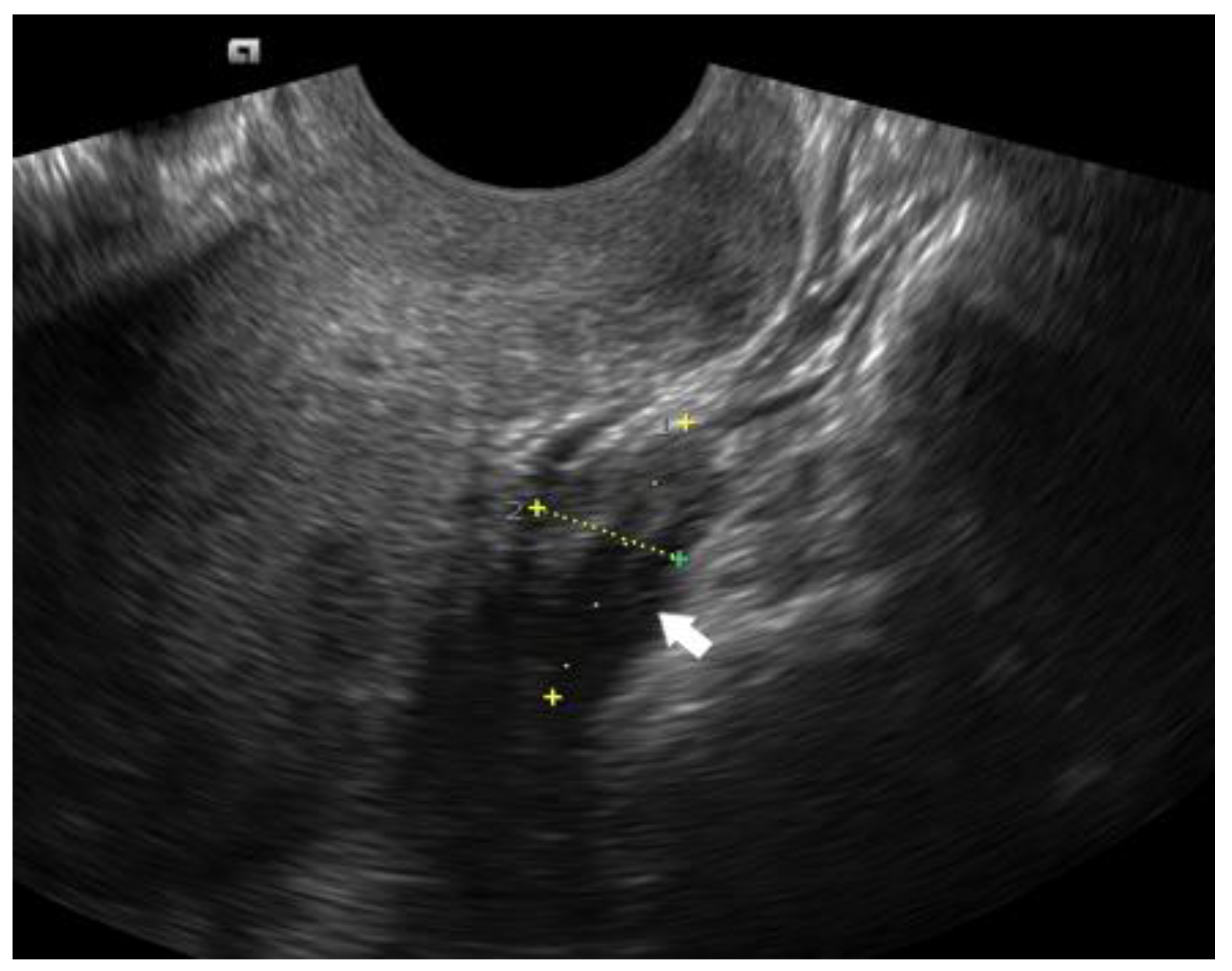
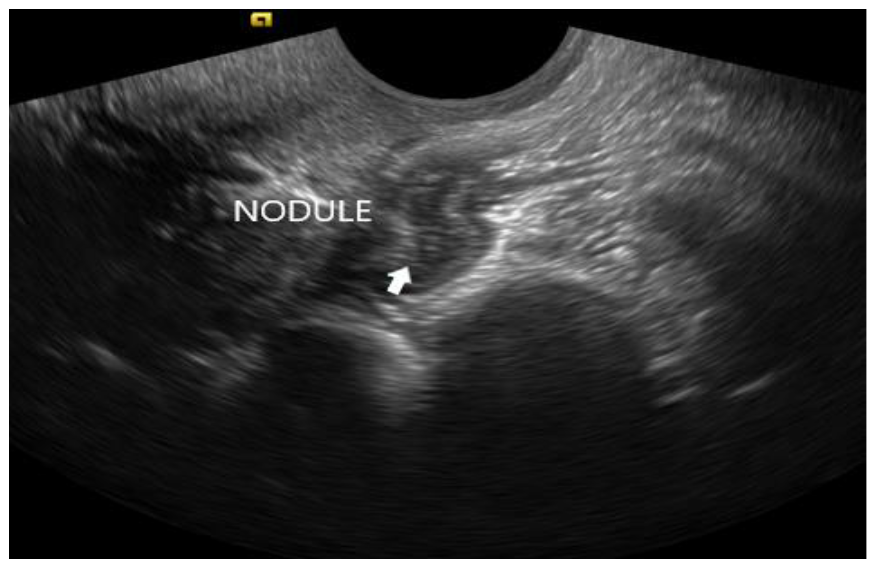
2.3. Deep Infiltrating Endometriosis (DIE)
2.4. The International Deep Endometriosis Analysis (IDEA) Approach
2.5. Anterior Compartment
2.5.1. Bladder
2.5.2. Ureter
2.6. Posterior Compartment
2.6.1. Torus Uterinus, Uterosacral Ligaments, Parametrium, Vagina, and Rectovaginal Septum (RVS)
2.6.2. Rectum and Sigmoid Colon
2.7. Other Ultrasound Techniques
2.8. Comparing TVS to MRI in the Assessment of DIE
2.9. Staging of Endometriosis
3. Conclusions
Author Contributions
Funding
Institutional Review Board Statement
Informed Consent Statement
Data Availability Statement
Conflicts of Interest
References
- Giudice, L.C.; Kao, L.C. Endometriosis. Lancet 2004, 364, 1789–1799. [Google Scholar] [CrossRef] [PubMed]
- Zondervan, K.T.; Becker, C.M.; Koga, K.; Missmer, S.A.; Taylor, R.N.; Viganò, P. Endometriosis. Nat. Rev. Dis. Primers 2018, 4, 9. [Google Scholar] [CrossRef] [PubMed]
- Bulun, S.E. Endometriosis. N. Engl. J. Med. 2009, 360, 268–279. [Google Scholar] [CrossRef]
- Leyendecker, G.; Bilgicyildirim, A.; Inacker, M.; Stalf, T.; Huppert, P.; Mall, G.; Böttcher, B.; Wildt, L. Adenomyosis and endometriosis. Re-visiting their association and further insights into the mechanisms of auto-traumatisation. An MRI study. Arch. Gynecol. Obstet. 2015, 291, 917–932. [Google Scholar] [CrossRef] [PubMed] [Green Version]
- Hudelist, G.; Fritzer, N.; Thomas, A.; Niehues, C.; Oppelt, P.; Haas, D.; Tammaa, A.; Salzer, H. Diagnostic delay for endometriosis in Austria and Germany: Causes and possible consequences. Hum. Reprod. 2012, 27, 3412–3416. [Google Scholar] [CrossRef] [Green Version]
- Nisenblat, V.; Bossuyt, P.M.M.; Farquhar, C.; Johnson, N.; Hull, M.L. Imaging modalities for the non-invasive diagnosis of endometriosis. Cochrane Database Syst. Rev. 2016, 2, CD009591. [Google Scholar] [CrossRef] [PubMed]
- Exacoustos, C.; Manganaro, L.; Zupi, E. Imaging for the evaluation of endometriosis and adenomyosis. Best Pract. Res. Clin. Obstet. Gynaecol. 2014, 28, 655–681. [Google Scholar] [CrossRef] [PubMed] [Green Version]
- Turocy, J.M.; Benacerraf, B.R. Transvaginal sonography in the diagnosis of deep infiltrating endometriosis: A review. J. Clin. Ultrasound 2017, 45, 313–318. [Google Scholar] [CrossRef]
- Van den Bosch, T.; Van Schoubroeck, D. Ultrasound diagnosis of endometriosis and adenomyosis: State of the art. Best Pract. Res. Clin. Obstet. Gynaecol. 2018, 51, 16–24. [Google Scholar] [CrossRef]
- Scioscia, M.; Virgilio, B.A.; Laganà, A.S.; Bernardini, T.; Fattizzi, N.; Neri, M.; Guerriero, S. Differential Diagnosis of Endometriosis by Ultrasound: A Rising Challenge. Diagnostics 2020, 10, 848. [Google Scholar] [CrossRef]
- Busacca, M.; Vignali, M. Ovarian endometriosis: From pathogenesis to surgical treatment. Curr. Opin. Obstet. Gynecol. 2003, 15, 321–326. [Google Scholar] [CrossRef] [PubMed]
- Redwine, D.B. Ovarian endometriosis: A marker for more extensive pelvic and intestinal disease. Fertil. Steril. 1999, 72, 310–315. [Google Scholar] [CrossRef] [PubMed]
- Van Holsbeke, C.; Van Calster, B.; Guerriero, S.; Savelli, L.; Paladini, D.; Lissoni, A.A.; Czekierdowski, A.; Fischerová, D.; Zhang, J.; Mestdagh, G.; et al. Endometriomas: Their ultrasound characteristics. Ultrasound Obstet. Gynecol. 2010, 35, 730–740. [Google Scholar] [CrossRef] [PubMed]
- Menakaya, U.; Reid, S.; Lü, C.; Bassem, G.; Infante, F.; Condous, G. Performance of ultrasound-based endometriosis staging system (UBESS) for predicting level of complexity of laparoscopic surgery for endometriosis. Ultrasound Obstet. Gynecol. 2016, 48, 786–795. [Google Scholar] [CrossRef] [Green Version]
- Guerriero, S.; Ajossa, S.; Mais, V.; Risalvato, A.; Lai, M.P.; Melis, G.B. The diagnosis of endometriomas using colour Doppler energy imaging. Hum. Reprod. 1998, 13, 1691–1695. [Google Scholar] [CrossRef] [Green Version]
- Timmerman, D.; Valentin, L.; Bourne, T.; Collins, W.P.; Verrelst, H.; Vergote, I. Terms, definitions and measurements to describe the sonographic features of adnexal tumors: A consensus opinion from the International Ovarian Tumor Analysis (IOTA) group. Ultrasound Obstet. Gynecol. 2000, 16, 500–505. [Google Scholar] [CrossRef] [Green Version]
- Valentin, L. Pattern recognition of pelvic masses by gray-scale ultrasound imaging: The contribution of Doppler ultrasound. Ultrasound Obstet. Gynecol. 1999, 14, 338–347. [Google Scholar] [CrossRef]
- Guerriero, S.; Condous, G.; van den Bosch, T.; Valentin, L.; Leone, F.P.G.; Van Schoubroeck, D.; Exacoustos, C.; Installé, A.J.F.; Martins, W.P.; Abrao, M.S.; et al. Systematic approach to sonographic evaluation of the pelvis in women with suspected endometriosis, including terms, definitions and measurements: A consensus opinion from the International Deep Endometriosis Analysis (IDEA) group. Ultrasound Obstet. Gynecol. 2016, 48, 318–332. [Google Scholar] [CrossRef]
- Mascilini, F.; Moruzzi, C.; Giansiracusa, C.; Guastafierro, F.; Savelli, L.; De Meis, L.; Epstein, E.; Timor-Tritsch, I.; Mailath-Pokorny, M.; Ercoli, A.; et al. Imaging in gynecological disease (10): Clinical and ultrasound characteristics of decidualized endometriomas surgically removed during pregnancy. Ultrasound Obstet. Gynecol. 2014, 44, 354–360. [Google Scholar] [CrossRef]
- Van Gorp, T.; Amant, F.; Neven, P.; Vergote, I.; Moerman, P. Endometriosis and the development of malignant tumours of the pelvis. A review of literature. Best Pract. Res. Clin. Obstet. Gynaecol. 2004, 18, 349–371. [Google Scholar] [CrossRef]
- Leonardi, M.; Robledo, K.; Espada, M.; Vanza, K.; Condous, G. SonoPODography: A new diagnostic technique for visualizing superficial endometriosis. Eur. J. Obstet. Gynecol. Reprod. Biol. 2020, 254, 124–131. [Google Scholar] [CrossRef] [PubMed]
- Koninckx, P.R.; Ussia, A.; Adamyan, L.; Wattiez, A.; Donnez, J. Deep endometriosis: Definition, diagnosis, and treatment. Fertil. Steril. 2012, 98, 564–571. [Google Scholar] [CrossRef] [PubMed]
- Guerriero, S.; Ajossa, S.; Orozco, R.; Perniciano, M.; Jurado, M.; Melis, G.B.; Alcazar, J.L. Accuracy of transvaginal ultrasound for diagnosis of deep endometriosis in the rectosigmoid: Systematic review and meta-analysis. Ultrasound Obstet. Gynecol. 2016, 47, 281–289. [Google Scholar] [CrossRef] [PubMed] [Green Version]
- Guerriero, S.; Ajossa, S.; Minguez, J.A.; Jurado, M.; Mais, V.; Melis, G.B.; Alcazar, J.L. Accuracy of transvaginal ultrasound for diagnosis of deep endometriosis in uterosacral ligaments, rectovaginal septum, vagina and bladder: Systematic review and meta-analysis. Ultrasound Obstet. Gynecol. 2015, 46, 534–545. [Google Scholar] [CrossRef] [PubMed]
- Hudelist, G.; English, J.; Thomas, A.E.; Tinelli, A.; Singer, C.F.; Keckstein, J. Diagnostic accuracy of transvaginal ultrasound for non-invasive diagnosis of bowel endometriosis: Systematic review and meta-analysis. Ultrasound Obstet. Gynecol. 2011, 37, 257–263. [Google Scholar] [CrossRef]
- Goncalves, M.O.; Neto, J.S.; Andres, M.P.; Siufi, D.; de Mattos, L.A.; Abrao, M.S. Systematic evaluation of endometriosis by transvaginal ultrasound can accurately replace diagnostic laparoscopy, mainly for deep and ovarian endometriosis. Hum. Reprod. 2021, 36, 1492–1500. [Google Scholar] [CrossRef]
- Indrielle-Kelly, T.; Frühauf, F.; Fanta, M.; Burgetova, A.; Lavu, D.; Dundr, P.; Cibula, D.; Fischerova, D. Diagnostic Accuracy of Ultrasound and MRI in the Mapping of Deep Pelvic Endometriosis Using the International Deep Endometriosis Analysis (IDEA) Consensus. BioMed Res. Int. 2020, 2020, 3583989. [Google Scholar] [CrossRef]
- Leonardi, M.; Uzuner, C.; Mestdagh, W.; Lu, C.; Guerriero, S.; Zajicek, M.; Dueckelmann, A.; Filippi, F.; Buonomo, F.; Pascual, M.A.; et al. Diagnostic accuracy of transvaginal ultrasound for detection of endometriosis using International Deep Endometriosis Analysis ( IDEA ) approach: Prospective international pilot study. Ultrasound Obstet. Gynecol. 2022, 60, 404–413. [Google Scholar] [CrossRef]
- Maggiore, U.L.R.; Ferrero, S.; Candiani, M.; Somigliana, E.; Vigano, P.; Vercellini, P. Bladder Endometriosis: A Systematic Review of Pathogenesis, Diagnosis, Treatment, Impact on Fertility, and Risk of Malignant Transformation. Eur. Urol. 2017, 71, 790–807. [Google Scholar] [CrossRef] [Green Version]
- Savelli, L.; Manuzzi, L.; Pollastri, P.; Mabrouk, M.; Seracchioli, R.; Venturoli, S. Diagnostic accuracy and potential limitations of transvaginal sonography for bladder endometriosis. Ultrasound Obstet. Gynecol. 2009, 34, 595–600. [Google Scholar] [CrossRef]
- Seracchioli, R.; Mabrouk, M.; Montanari, G.; Manuzzi, L.; Concetti, S.; Venturoli, S. Conservative laparoscopic management of urinary tract endometriosis (UTE): Surgical outcome and long-term follow-up. Fertil. Steril. 2010, 94, 856–861. [Google Scholar] [CrossRef]
- Chapron, C.; Chiodo, I.; Leconte, M.; Amsellem-Ouazana, D.; Chopin, N.; Borghese, B.; Dousset, B. Severe ureteral endometriosis: The intrinsic type is not so rare after complete surgical exeresis of deep endometriotic lesions. Fertil. Steril. 2010, 93, 2115–2120. [Google Scholar] [CrossRef] [PubMed]
- Nezhat, C.; Paka, C.; Gomaa, M.; Schipper, E. Silent Loss of Kidney Seconary to Ureteral Endometriosis. J. Soc. Laparoendosc. Surg. 2012, 16, 451–455. [Google Scholar] [CrossRef] [PubMed] [Green Version]
- Savelli, L.; Ambrosio, M.; Salucci, P.; Raimondo, D.; Arena, A.; Seracchioli, R. Transvaginal ultrasound features of normal uterosacral ligaments. Fertil. Steril. 2021, 116, 275–277. [Google Scholar] [CrossRef] [PubMed]
- Bazot, M.; Lafont, C.; Rouzier, R.; Roseau, G.; Thomassin-Naggara, I.; Daraï, E. Diagnostic accuracy of physical examination, transvaginal sonography, rectal endoscopic sonography, and magnetic resonance imaging to diagnose deep infiltrating endometriosis. Fertil. Steril. 2009, 92, 1825–1833. [Google Scholar] [CrossRef] [PubMed]
- Reid, S.; Lu, C.; Casikar, I.; Reid, G.; Abbott, J.; Cario, G.; Chou, D.; Kowalski, D.; Cooper, M.; Condous, G. Prediction of pouch of Douglas obliteration in women with suspected endometriosis using a new real-time dynamic transvaginal ultrasound technique: The sliding sign. Ultrasound Obstet. Gynecol. 2012, 41, 685–691. [Google Scholar] [CrossRef] [PubMed]
- Scarella, A.C.; Devoto, L.C.; Villarroel, C.Q.; Inzunza, N.P.; Quilodrán, F.R.; Sovino, H.S. Transvaginal ultrasound for preoperative detection of deep endometriosis in patients with chronic pelvic pain [Ultrasonido transvaginal para la detección preoperatoria de endometriosis profunda en pacientes con dolor pélvico crónico]. Rev. Chil. Obs. Ginecol. 2013, 78, 114–118. [Google Scholar] [CrossRef] [Green Version]
- Bazot, M.; Thomassin, I.; Hourani, R.; Cortez, A.; Darai, E. Diagnostic accuracy of transvaginal sonography for deep pelvic endometriosis. Ultrasound Obstet. Gynecol. 2004, 24, 180–185. [Google Scholar] [CrossRef] [PubMed]
- Guerriero, S.; Ajossa, S.; Gerada, M.; Virgilio, B.; Angioni, S.; Melis, G.B. Diagnostic value of transvaginal ‘tenderness-guided’ ultrasonography for the prediction of location of deep endometriosis. Hum. Reprod. 2008, 23, 2452–2457. [Google Scholar] [CrossRef] [Green Version]
- Abo, C.; Moatassim, S.; Marty, N.; Ghislain, M.S.; Huet, E.; Bridoux, V.; Tuech, J.J.; Roman, H. Postoperative complications after bowel endometriosis surgery by shaving, disc excision, or segmental resection: A three-arm comparative analysis of 364 consecutive cases. Fertil. Steril. 2018, 109, 172–178.e1. [Google Scholar] [CrossRef]
- Roman, H.; Ness, J.; Suciu, N.; Bridoux, V.; Gourcerol, G.; Leroi, A.M.; Tuech, J.J.; Ducrotté, P.; Savoye-Collet, C.; Savoye, G. Are digestive symptoms in women presenting with pelvic endometriosis specific to lesion localizations? A preliminary prospective study. Hum. Reprod. 2012, 27, 3440–3449. [Google Scholar] [CrossRef] [PubMed]
- Roman, H.; Bubenheim, M.; Huet, E.; Bridoux, V.; Zacharopoulou, C.; Daraï, E.; Collinet, P.; Tuech, J.-J. Conservative surgery versus colorectal resection in deep endometriosis infiltrating the rectum: A randomized trial. Hum. Reprod. 2018, 33, 47–57. [Google Scholar] [CrossRef] [PubMed]
- Gerges, B.; Li, W.; Leonardi, M.; Mol, B.W.; Condous, G. Optimal imaging modality for detection of rectosigmoid deep endometriosis: Systematic review and meta-analysis. Ultrasound Obstet. Gynecol. 2021, 58, 190–200. [Google Scholar] [CrossRef] [PubMed]
- Piessens, S.; Healey, M.; Maher, P.; Tsaltas, J.; Rombauts, L. Can anyone screen for deep infiltrating endometriosis with transvaginal ultrasound? Aust. N. Z. J. Obstet. Gynaecol. 2014, 54, 462–468. [Google Scholar] [CrossRef] [PubMed]
- Tammaa, A.; Fritzer, N.; Strunk, G.; Krell, A.; Salzer, H.; Hudelist, G. Learning curve for the detection of pouch of Douglas obliteration and deep infiltrating endometriosis of the rectum. Hum. Reprod. 2014, 29, 1199–1204. [Google Scholar] [CrossRef] [PubMed]
- Deslandes, A.; Parange, N.; Childs, J.T.; Osborne, B.; Panuccio, C.; Croft, A.; Bezak, E. How long does a transvaginal ultrasound examination for endometriosis take in comparison to a routine transvaginal ultrasound examination? Australas. J. Ultrasound Med. 2021, 25, 20–27. [Google Scholar] [CrossRef]
- De Ceglie, A.; Bilardi, C.; Blanchi, S.; Picasso, M.; Di Muzio, M.; Trimarchi, A.; Conio, M. Acute small bowel obstruction caused by endometriosis: A case report and review of the literature. World J. Gastroenterol. 2008, 14, 3430–3434. [Google Scholar] [CrossRef]
- Guerriero, S.; Pascual, M.A.; Ajossa, S.; Rodríguez, I.; Zajicek, M.; Rolla, M.; Llop, N.R.; Yulzari, V.; Bardin, R.; Buonomo, F.; et al. Learning curve for ultrasonographic diagnosis of deep infiltrating endometriosis using structured offline training program. Ultrasound Obstet. Gynecol. 2019, 54, 262–269. [Google Scholar] [CrossRef]
- Reid, S.; Lu, C.; Hardy, N.; Casikar, I.; Reid, G.; Cario, G.; Chou, D.; Almashat, D.; Condous, G. Office gel sonovaginography for the prediction of posterior deep infiltrating endometriosis: A multicenter prospective observational study. Ultrasound Obstet. Gynecol. 2014, 44, 710–718. [Google Scholar] [CrossRef]
- Dessole, S.; Farina, M.; Rubattu, G.; Cosmi, E.; Ambrosini, G.; Nardelli, G.B. Sonovaginography is a new technique for assessing rectovaginal endometriosis. Fertil. Steril. 2003, 79, 1023–1027. [Google Scholar] [CrossRef]
- Bergamini, V.; Ghezzi, F.; Scarperi, S.; Raffaelli, R.; Cromi, A.; Franchi, M. Preoperative assessment of intestinal endometriosis: A comparison of Transvaginal Sonography with Water-Contrast in the Rectum, Transrectal Sonography, and Barium Enema. Gastrointest. Radiol. 2010, 35, 732–736. [Google Scholar] [CrossRef] [PubMed]
- Menada, M.V.; Remorgida, V.; Abbamonte, L.; Nicoletti, A.; Ragni, N.; Ferrero, S. Does transvaginal ultrasonography combined with water-contrast in the rectum aid in the diagnosis of rectovaginal endometriosis infiltrating the bowel? Hum. Reprod. 2008, 23, 1069–1075. [Google Scholar] [CrossRef]
- Morotti, M.; Ferrero, S.; Bogliolo, S.; Venturini, P.L.; Remorgida, V.; Menada, M.V. Transvaginal ultrasonography with water-contrast in the rectum in the diagnosis of bowel endometriosis. Minerva Ginecol. 2010, 62, 179–185. [Google Scholar] [PubMed]
- Bignardi, T.; Condous, G. Sonorectovaginography: A new sonographic technique for imaging of the posterior compartment of the pelvis. J. Ultrasound Med. 2008, 27, 1479–1483. [Google Scholar] [CrossRef] [PubMed]
- Bazot, M.; Detchev, R.; Cortez, A.; Amouyal, P.; Uzan, S.; Daraï, E. Transvaginal sonography and rectal endoscopic sonography for the assessment of pelvic endometriosis: A preliminary comparison. Hum. Reprod. 2003, 18, 1686–1692. [Google Scholar] [CrossRef] [Green Version]
- National Guideline Alliance (UK). Endometriosis: Diagnosis and Management; National Institute for Health and Care Excellence (NICE): London, UK, September 2017. [Google Scholar]
- Gerges, B.; Li, W.; Leonardi, M.; Mol, B.W.; Condous, G. Meta-analysis and systematic review to determine the optimal imaging modality for the detection of uterosacral ligaments/torus uterinus, rectovaginal septum and vaginal deep endometriosis. Hum. Reprod. Open 2021, 2021, hoab041. [Google Scholar] [CrossRef] [PubMed]
- Guerriero, S.; Saba, L.; Pascual, M.A.; Ajossa, S.; Rodriguez, I.; Mais, V.; Alcazar, J.L. Transvaginal ultrasound vs magnetic resonance imaging for diagnosing deep infiltrating endometriosis: Systematic review and meta-analysis. Ultrasound Obstet. Gynecol. 2018, 51, 586–595. [Google Scholar] [CrossRef] [Green Version]
- Gerges, B.; Li, W.; Leonardi, M.; Mol, B.; Condous, G. Meta-analysis and systematic review to determine the optimal imaging modality for the detection of bladder deep endometriosis. Eur. J. Obstet. Gynecol. Reprod. Biol. 2021, 261, 124–133. [Google Scholar] [CrossRef]
- Pereira, A.M.G.; Brizon, V.S.C.; Junior, N.C.; Carvalho, M.D.S.R.D.; Cuvero, E.M.; Buehler, A.M.; Pereira, T.D.V.; Lopes, R.G.C. Can Enhanced Techniques Improve the Diagnostic Accuracy of Transvaginal Sonography and Magnetic Resonance Imaging for Rectosigmoid Endometriosis? A Systematic Review and Meta-analysis. J. Obstet. Gynaecol. Can. 2020, 42, 488–499.e4. [Google Scholar] [CrossRef]
- Gutiérrez, A.H.; Spagnolo, E.; Hidalgo, P.; López, A.; Zapardiel, I.; Rodriguez, R. Magnetic resonance imaging versus transvaginal ultrasound for complete survey of the pelvic compartments among patients with deep infiltrating endometriosis. Int. J. Gynecol. Obstet. 2019, 146, 380–385. [Google Scholar] [CrossRef]
- Medeiros, L.R.; Rosa, M.I.; Silva, B.R.; Reis, M.E.; Simon, C.S.; Dondossola, E.R.; Filho, J.S.D.C. Accuracy of magnetic resonance in deeply infiltrating endometriosis: A systematic review and meta-analysis. Arch. Gynecol. Obstet. 2015, 291, 611–621. [Google Scholar] [CrossRef] [PubMed]
- Mason, B.R.; Chatterjee, D.; Menias, C.O.; Thaker, P.H.; Siegel, C.L.; Yano, M. Encyclopedia of endometriosis: A pictorial rad-path review. Abdom. Radiol. 2020, 45, 1587–1607. [Google Scholar] [CrossRef] [PubMed]
- Classification of endometriosis. The American Fertility Society. Fertil Steril. 1979, 32, 633–634. [Google Scholar]
- Canis, M.; Donnez, J.G.; Guzick, D.S.; Halme, J.K.; Rock, J.A.; Schenken, R.S.; Vernon, M.W. Revised American Society for Reproductive Medicine classification of endometriosis: 1996. Fertil. Steril. 1997, 67, 817–821. [Google Scholar] [CrossRef]
- Leonardi, M.; Espada, M.; Choi, S.; Chou, D.; Chang, T.; Smith, C.; Rowan, K.; Condous, G. Transvaginal Ultrasound Can Accurately Predict the American Society of Reproductive Medicine Stage of Endometriosis Assigned at Laparoscopy. J. Minim. Invasive Gynecol. 2020, 27, 1581–1587.e1. [Google Scholar] [CrossRef] [Green Version]
- Tuttlies, F.; Keckstein, J.; Ulrich, U.; Possover, M.; Schweppe, K.W.; Wustlich, M.; Buchweitz, O.; Greb, R.; Kandolf, O.; Mangold, R.; et al. ENZIAN-Score, eine Klassifikation der tief infiltrierenden Endometriose [ENZIAN-score, a classification of deep infiltrating endometriosis]. Zent. Gynakol. 2005, 127, 275–281. [Google Scholar] [CrossRef]
- Keckstein, J.; Saridogan, E.; Ulrich, U.A.; Sillem, M.; Oppelt, P.; Schweppe, K.W.; Krentel, H.; Janschek, E.; Exacoustos, C.; Malzoni, M.; et al. The #Enzian classification: A comprehensive non-invasive and surgical description system for endometriosis. Acta Obstet. Gynecol. Scand. 2021, 100, 1165–1175. [Google Scholar] [CrossRef]
- Hudelist, G.; Montanari, E.; Salama, M.; Dauser, B.; Nemeth, Z.; Keckstein, J. Comparison between Sonography-based and Surgical Extent of Deep Endometriosis Using the Enzian Classification—A Prospective Diagnostic Accuracy Study. J. Minim. Invasive Gynecol. 2021, 28, 1643–1649.e1. [Google Scholar] [CrossRef]
- Montanari, E.; Bokor, A.; Szabó, G.; Kondo, W.; Trippia, C.H.; Malzoni, M.; Di Giovanni, A.; Tinneberg, H.R.; Oberstein, A.; Rocha, R.M.; et al. Accuracy of sonography for non-invasive detection of ovarian and deep endometriosis using #Enzian classification: Prospective multicenter diagnostic accuracy study. Ultrasound Obstet. Gynecol. 2022, 59, 385–391. [Google Scholar] [CrossRef]
- Di Giovanni, A.; Montanari, E.; Hudelist, G.; Malzoni, M.; Keckstein, J. Comparison Between Sonography-Based and Surgical Evaluation of Endometriotic Lesions Using the #Enzian Classification—A Retrospective Data Analysis. Ultraschall Der Med.—Eur. J. Ultrasound 2022. [Google Scholar] [CrossRef]
- Espada, M.; Leonardi, M.; Aas-Eng, K.; Lu, C.; Reyftmann, L.; Tetstall, E.; Slusarczyk, B.; Ludlow, J.; Hudelist, G.; Reid, S.; et al. A Multicenter International Temporal and External Validation Study of the Ultrasound-based Endometriosis Staging System. J. Minim. Invasive Gynecol. 2021, 28, 57–62. [Google Scholar] [CrossRef] [PubMed]
- Chaabane, S.; Xuan, H.-T.N.; Paternostre, A.; Du Cheyron, J.; Harizi, R.; Mimouni, M.; Fauconnier, A. Endométriose: Évaluation du score Ultrasound-Based Endometriosis Staging System (UBESS) dans la prédiction de la difficulté chirurgicale [Endometriosis: Assessment of the Ultrasound-Based Endometriosis Staging System score (UBESS) in predicting surgical difficulty]. Gynecol Obs. Fertil Senol. 2019, 47, 265–272. [Google Scholar] [CrossRef]


Publisher’s Note: MDPI stays neutral with regard to jurisdictional claims in published maps and institutional affiliations. |
© 2022 by the authors. Licensee MDPI, Basel, Switzerland. This article is an open access article distributed under the terms and conditions of the Creative Commons Attribution (CC BY) license (https://creativecommons.org/licenses/by/4.0/).
Share and Cite
Daniilidis, A.; Grigoriadis, G.; Dalakoura, D.; D’Alterio, M.N.; Angioni, S.; Roman, H. Transvaginal Ultrasound in the Diagnosis and Assessment of Endometriosis—An Overview: How, Why, and When. Diagnostics 2022, 12, 2912. https://doi.org/10.3390/diagnostics12122912
Daniilidis A, Grigoriadis G, Dalakoura D, D’Alterio MN, Angioni S, Roman H. Transvaginal Ultrasound in the Diagnosis and Assessment of Endometriosis—An Overview: How, Why, and When. Diagnostics. 2022; 12(12):2912. https://doi.org/10.3390/diagnostics12122912
Chicago/Turabian StyleDaniilidis, Angelos, Georgios Grigoriadis, Dimitra Dalakoura, Maurizio N. D’Alterio, Stefano Angioni, and Horace Roman. 2022. "Transvaginal Ultrasound in the Diagnosis and Assessment of Endometriosis—An Overview: How, Why, and When" Diagnostics 12, no. 12: 2912. https://doi.org/10.3390/diagnostics12122912