General Topics (Dugesia, Schistosoma, fasciola, Taneia solium)
Classification of Animals Non Chordates of Class 11
Dugesia (Planaria)
- Fresh water form found in slow moving streams or fresh water ponds. The animal glides over solid submerged structures by cilia, mucilage and muscular contractions.
- It can also attach itself to the substratum by adhesive zone present ventrally.
- The body has a triangular anterior head with two lateral lobes or auricles & two black light sensitive dorsal eyes (no image is formed).
- Mouth lies ventrally. In the middle a small gonopore lies posterior to the mouth. The anus in absent.
- Planaria feeds on small crustaceans. The undigested food is passed out through the pharynx.
- Nephridiopores occur dorsally along the two sides.
- The animal can multiply asexually by binary fission. The power of regeneration is high.
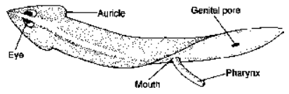
Fig. Flatworms : Turbellaria
Schistosoma (Blood fluke)
- The parasite lives in the hepatic portal system & mesentric blood vessels of human beings (Primary host).
- The secondary host is snail.
- Life cycle includes larval stages such as miracidium, sporocyst, cercaria larva. Cercaria larva directly penetrates the human skin.
- Its infection causes itching, rashes, body aches, fever, eosinophilia & may lead to congestive heart failure. It causes disease called schistosomiasis.
- It exhibits sexual dimorphism, the female is longer than male.
- Male & female have oral sucker & acetabulum (ventral sucker) but in male acetabulum is larger & more powerful.
- The female blood fluke resides in the gynecophoral canal of male.
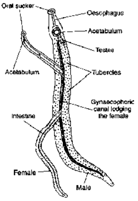
Fig. Schistosoma : Male and female
Fasciola (Liver fluke)
- Digestive, endoparasite found in the bile duct of the liver of sheep & goat which serve as primary host & in larval stages in an intermediate (secondary) host called Limnea (snail).
- In primary host it causes liver rot.
- The anterior end of the body is the form of a conical projection, called the head lobe at the tip of which oral sucker having mouth is present.
- There are present two muscular suckers; an oral sucker enclosing a mouth, and a ventral sucker or acetabulum without an aperture which is placed ventrally. The suckers are organs for attachment although anterior sucker also helps in taking the food.
- There are three permanent apertures on the body-mouth, common genital pore and excretory pore.
- With the help of mouth, the fluke feeds on bile, blood, lymph and cell debris of the host. There is no anus.
- Indigestible food matter is either probably ejected through the mouth or diffused into the excretory system.
- Respiration takes place in the absence of oxygen.
- The fluke is hermaphrodite but there is cross fertilization in bile duct of sheep.

Fig. Flatworms : Trematoda
Life History
- Cross fertilization takes place. The everted cirrus of one fluke penetrates the Laurer’s canal of the other through the opening & injects spermatozoa into the oviduct.
- A single fluke may produce a total of about 200,000 eggs in about 11 years.Each fertilized egg is surrounded by yolk cells, which provide yolk & shell material. The encapsulated embryo does not develop further in uterus. It is finally ejected out with faeces of host.
- When suitable conditions commence then encapsulated embryo differentiates into a miracidium larva. It swims in the water and is free living. It does not feed.
- When mirocidium comes in contact with intermediate snail host it enters the digestive gland. It develop into the second larva i.e., sporocyst larva.
- The sporocyst moves in the tissues of the host, absorbing nutrition from it. Each sporocyst produces five to eight rediae.
- Rediae emerges out from sporocyst and feeds upon host’s tissue.
- Cercaria larva is formed in the body of redia larva. It leaves the body of redia & ultimately escape the host body in surrounding water.
- The cercaria swims in surrounding water and later on becomes inactive to form metacercaria larva. This larva is encysted, so when primary host feeds on grasses, it enters in the body and establishes itself in the liver, where it becomes mature.
- Fasciola is supposed to show the phenomenon of polyembryony (larval stages give birth to next larval stages).
Parasitic adaptations of Fasciola
- A protective tegument around body is present.
- Simple digestive system without digestive glands because adult take almost digested food.
- Simple nervous system.
- Locomotary organs are absent in adult.
- Sense organs are absent in adult, eyes present only in larval stage.
- Spines and scales are present to prevent fluke from being washed down the bile ducts of host and for eroding tissues of host for feeding.
- Suckers are present for attachment.
- Enormously developed reproductive system.
- Reproduction in larval stages facilitate increase in number.

Fig. Life Cycle of Fasciola
Taenia solium
A pork tapeworm is ribbon-shaped, digenetic endoparasite.
The adults of Taenia solium are parasites in the small intesine of human beings (Primary host) & its larva infests the muscles of the pig (secondary host).
The body is divisible into three parts :
Scolex : At the apex is a prominent mobile cone, the rostellum which is armed with 22 to 32 curved chitinous hooks, arranged in two rows around its base, one row is of larger hooks & other row is of small
hooks. Both types of hooks alternate with each other. The scolex also bears four hemispherical, highly muscular suctorial organs, the true suckers. The suckers are devoid of hooks or spines. The suckers &
hooks are the organs of attachment. They don’t play any role in catching food.
Neck : It is a small narrow undifferentiated part behind the scolex. Its hinder end (area of segmentation) adds new segment to strobila.
Strobila : It consists of 800-1,000 segments or proglottids. The anterior proglottids are immature & have no reproductive organs; from about the 200th proglottids, the male organs are formed. From about 250th
to 650th proglottids, there are both sex organs; these are mature proglottids. The remaining proglottids lose the sex organs & have a distended uterus filled with eggs; these are called gravid proglottids. Last
gravid segment usually detach one by one from the body & pass out with the faeces of the host-the phenomenon is known as Apolysis.
Body wall : The outermost covering of the body is the cuticle. Below cuticle lies a basement membrane which separates the muscular layer and parenchyma from the cuticle. In the subcuticular region there are
circular and longitudinal muscles. The parenchyma is made up of loosely arranged parenchymatous cells. Gland cells are also found in between muscles. These gland cells secrete the protective secretion against the digestive enzymes.
Digestive system is totally absent; they absorb nutrients through their body wall. Respiration is anaerobic; excretion is through flame cells.
Reproductive system :
Male organ include numerous testes of many small lobes lying scattered in the greater part of the proglottids towards the dorsal side; efferent ducts arise from the testes & unite to open into a convoluted vas deferens which passes through a cirrus surrounded by cirrus sac. The cirrus opens by the male genital pore.
Female reproductive organs include a single, bilobed ovary; single oviduct opens into ootype; ootype surrounded by mehlis glands, vagina combines ootype with common genital atrium & opens outside by female genital aperture; forms seminal receptacle before opening outside; long, tubular blind sac-like uterus connected with ootype.
Life History
- The process of fertilization takes place either within the same proglottid or between two different proglottids to form zygote.
- The zygotes pass into the ootype, where they are enclosed by thick capsule. This capsulated zygote then passes into the uterus. Passage of capsules into the uterus is lubricated by the secretion from mehlis glands.
- The process of cleavage takes place to form morula. The morula at posterior end develops three pairs of chitinous hooks. The six-hooked embryo is called hexacanth.
- The hexacanth is surrounded by two membranes. This hexacanth alongwith the membrane is known as onchosphere. Onchosphere passes out with stool by process of apolysis (shedding of gravid proglottid).
- The secondary host acquires infection by ingesting the onchospheres. (Man may act as secondary host if detached gravid proglottids are passd into intestine by reverse peristalsis.)
- The onchosphere looses its membrane by the action of gastric enzymes. The hexacanth then passes into the small intestine. The hexacanth is now activated by bile salts. It reaches muscles (striated) by boring through intestinal mucosa. It now forms encysted bladderworm or cysticercus larva.
- The cysticercus develops in the adult tapeworm only when ingested by the human beings. Pork (Pig’s flesh) containing cysticerci is called measly pork for its spotted appearance.
- The human host gets the infection by eating under-cooked measly pork.
- Cysticercosis is more harmful than taeniasis.
Taenia saginata
It commonly known as beef tapeworm where the intermediate host is cow, buffalo and sheep. Rostellum hooks are absent. It is longer than T. solium and it is the commonest tape worm.
Echinococcus granulosus : It is commonly known as dog tapeworm or hydatid tapeworm. It is an endoparasite in dogs, cats, fox and other carnivores. It is the smallest tapeworm. It has well developed scolex
with four suckers & rostellum with a double row of hooks.
Parasitic Adaptation in flatworms
They are well adapted for anaerobic respiration
They are mostly ehrmaphrodite which ensures copulation. They also produce large number of eggs.
They also protect themselves by secreting mucous & antienzymes. Antienzymes neutralise digestive enzymes.
Organ of adhesion : They have well-developed suckers, hooks and spines.
Organ of nutrition : They feed on readily available digested & semi-digested food of the host. Digestive system totally absent in cestode & poorly developed in trematoda.
Body covering - made of thick tegument.
- Introduction
- Fundamentals of Animal Classification
- Body Plan and Symmetry
- Metamerism and Germ Layers
- Body Cavity or Coelom
- Protostomes And Deuterostomes
- Modern Classification of Animal World
- Five Kingdoms of Living World
- Kingdom Protista
- Protozoa
- Plasmodium
- Some Representatives of Phylum Protozoa
- Paramecium
- Phylum Porifera
- Canal System and Skeleton
- Some representatives of phylum - Porifera
- Cnidaria (Gr. Knide = nettle or stringing cell)
- Some representatives of phylum Coelentrata
- Phylum Platyhelminthes - The flat worms
- General Topics (Dugesia, Schistosoma, fasciola, Taneia solium)
- Important Points to Remember
- Phylum Aschelminthes
- Some representatives of Nematoda
- Phylum Annelida
- Polychaeta
- Oligochaeta
- Hirudinea
- Phylum Arthoropoda
- General Topics
- Important Points to Remember of Chelicerae
- Phylum Mollusca
- General Topics of Phylum Mollusca
- Important Points to Remember of Phylum Mollusca
- Phylum Echinodermata
- General Topics of Phylum Echinodermata
- Important Points to Remember of Phylum Echinodermata
- Exercise 1
- Exercise 2
- Exercise 3
- Exercise 4
- Exercise 5
- Exercise 6