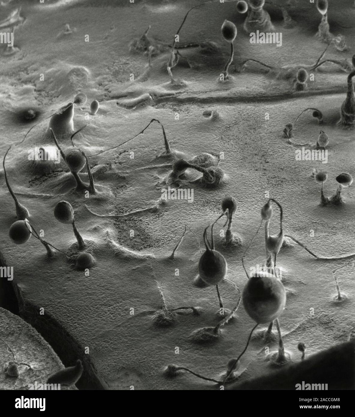
Scanning electron micrograph of spore towers of the slime mould Dictyostelium discoideum. Slime moulds are a curious division of plants with many of t
RMID:Image ID:2ACCGM8
Image details
Contributor:
Science Photo Library / Alamy Stock PhotoImage ID:
2ACCGM8File size:
33 MB (1.5 MB Compressed download)Releases:
Model - no | Property - noDo I need a release?Dimensions:
3241 x 3561 px | 27.4 x 30.1 cm | 10.8 x 11.9 inches | 300dpiDate taken:
5 March 1986Photographer:
DAVID SCHARF/SCIENCE PHOTO LIBRARYMore information:
Scanning electron micrograph of spore towers of the slime mould Dictyostelium discoideum. Slime moulds are a curious division of plants with many of the characteristics of animals. They consist of a slow-moving mass of amoeboid protoplasm, which engulfs particles of solid food which it encounters. It reproduces itself however by spores, shed from a spore tower, produced by a vegetative body called the plasmodium. The outer slime sheath that surrounds the top of each tower disappears with maturity, exposing the spores. The majority of slime moulds are saprophytes, feeding on dead or decaying organic matter. Magnification: X 50 (at 10x8 size).